Atrial Flutter Ablation Patient Information
What is atrial flutter?
Atrial flutter refers to an abnormal heart rhythm involving an electrical short circuit occurring in the heart’s upper chambers.
How does the heart normally work?
The heart as a pump
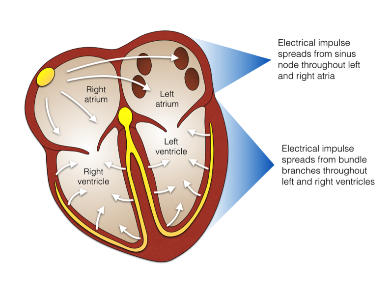
The heart is a muscular pump that circulates blood around the body. The right side of the heart has 2 chambers: the right atrium and the right ventricle. The right atrium receives blood as it returns from the body and channels it through a valve into the right ventricle. The right ventricle then pumps blood to the lungs where it collects oxygen to deliver to the rest of the body. The left side of the heart also has 2 chambers: the left atrium and the left ventricle. The left atrium receives blood from the lungs and channels it through another valve into the left ventricle. The left ventricle then pumps blood to the rest of the body via the arteries. The left and right atria are positioned side by side and form the heart’s upper chambers, while the left and right ventricles form the lower chambers.

The heart’s electrical system
The heart has an electrical system that co-ordinates its pump function. Every normal heartbeat starts with an electrical impulse generated by the heart’s own pacemaker or “spark plug”, called the sino-atrial node (SA node). The SA node sets the heart’s rhythm and rate by firing 60 to 100 times a minute. It fires more rapidly during exercise and stress, and slows down during rest or sleep.
Each electrical impulse from the SA node spreads through the right and left atria like a ripple in a pond, causing the chambers to contract. The impulse reaches the atrio-ventricular node (AV node) in the centre of the heart, and is then transmitted into the lower chambers via specialized electrical cables called “bundle branches” that course through the right and left ventricles. The AV node is a critical structure in the heart as it is normally forms the only electrical connection between the upper and lower chambers. As the electrical impulse is delivered to the ventricles, it causes them to contract and to pump blood to the lungs and the body.
The SA node, the AV node, and the specialized electrical cables of the lower chambers of the heart need to function correctly to generate the normal heartbeat. When the normal sequence of electrical activity is disrupted, an abnormal heart rhythm will result.
How does atrial flutter occur?
Atrial flutter typically refers to an electrical short circuit involving the right atrium which cycles at around 300 times per minute. The short circuit is triggered by ectopic beats or “electrical sparks” usually arising from the left atrium. During atrial flutter, both the left and right atria cannot pump effectively at this very rapid rate. The AV node will normally allow only half of these electrical impulses to reach the heart’s lower pumping chambers, resulting in a pulse rate of 150 beats per minute. Medications including beta blockers (metoprolol, atenolol, etc), calcium channel blockers (verapamil, diltiazem), and digoxin help the AV node filter out more of these rapid impulses to bring the heart rate into a normal range, even while the upper chambers remain in short circuit.

There are 3 important consequences of atrial flutter:
- The heart becomes inefficient at pumping blood around the body. This produces breathlessness, dizziness, and fatigue, often accompanied by palpitations. These are the key symptoms of atrial flutter, and are not in themselves life threatening.
- Blood flow in the heart’s upper chambers slows down, and can stagnate in the “nooks and crannies” of the left atrium. This sometimes results in formation of blood clots which if released into circulation can cause stroke. Depending on a number of factors, many patients will require a blood thinner to prevent blood clot formation and stroke. This may be warfarin, or one of the new generation of blood thinners: Pradaxa, Xarelto, or Eliquis.
- Atrial flutter causes the heart’s lower chambers to pump too quickly. If left untreated, these muscular chambers can weaken and distend, resulting in heart failure. It is important to prevent this either by slowing down the irregular heart rate with medications, or correcting the short circuit altogether so that normal electrical function resumes.
How is atrial flutter treated?
There are 3 main options for people with atrial flutter.
- Medications. Atrial flutter can sometimes be managed with medications. However, in many individuals, medications are ineffective or cause unwanted side effects. Your doctor will discuss the various options with you including their various risks and benefits.
- Electrical cardioversion. Atrial flutter can be terminated with an electrical shock to the chest, delivered while you are under a very brief period of general anaesthesia. This “resets” the heart’s electrical system, allowing normal rhythm to resume. This strategy is almost always successful acutely, however there is a 50% chance of developing atrial flutter again in the next 12 months. Most patients will require an antiarrhythmic medication such as flecainide, sotalol, or amiodarone to prevent atrial flutter from returning.
- Catheter ablation. This is a procedure that cures the condition by using a keyhole instrument called a “catheter”. The catheter is used to deliver radiofrequency energy to ablate (destroy) a critical part of the short circuit in the right atrium which breaks the circuit and prevents it from recurring. The success rate is 95%.

What is catheter ablation?
A catheter is a fine wire that can be threaded through a blood vessel via a keyhole opening and placed in the heart. Usually, a blood vessel in the top of the right thigh in the groin area is used for access to the heart. Each catheter has electrodes on its end that can monitor the heart’s internal electrical activity.
A special “ablation catheter” can be used to deliver radiofrequency energy to the problem area in the heart. Radiofrequency energy is a low power, high frequency energy that causes a tiny region of the heart near the tip of the catheter to increase in temperature, thus ablating (or cauterising) a small area of tissue.
Radiofrequency energy has been used for decades by surgeons to cut tissue or to stop bleeding. For the treatment of palpitations, a much lower power of radio-frequency energy is used.
What happens prior to the procedure?
If you are taking a blood thinner for your heart condiction, you will need to continue taking this as usual. You will normally be admitted on the day of your procedure. Prior to the procedure you will normally have an ECG and blood test. You will be required to fast for at least six hours before the study.
If your procedure is in the afternoon, you may have a light early breakfast. If your procedure is in the morning, DO NOT EAT OR DRINK AFTER MIDNIGHT, except for sips of water to help you swallow your pills.
What happens during a catheter ablation procedure?
You will be transferred to the Electrophysiology Laboratory (EP lab) from your ward or from the waiting area. Usually before leaving you will be given a light sedative and your groin will be shaved. This is because 3 small tubes will need to be placed in the blood vessel at the top of the right thigh in the groin area during the EP study.
The EP lab has a patient table, X-Ray tube, ECG monitors and various equipment. The staff in the lab will all be dressed in hospital theater clothes and during the procedure will be wearing hats and masks. Many ECG monitoring electrodes will be attached to your chest area and patches to your chest and back. These patches may momentarily feel cool on your skin.
A nurse or doctor will insert an intravenous line usually into the back of your hand. This is needed as a reliable way to give you medications during the study without further injections. You will also be given further sedation if and as required. You will also have a blood-pressure cuff attached to your arm that will automatically inflate at various times throughout the procedure.
The oxygen level of your blood will also be measured during the EP study and a small plastic device will be fitted on your finger for this purpose. Your groin area and possibly your neck will be washed with an antiseptic cleansing liquid and you will be covered with sterile sheets leaving these areas exposed.
An anesthetist will be present for many procedures. The procedure may be performed under local anaesthetic with sedative medication or under full general anaesthetic. This will be discussed with you before the procedure. If the procedure is performed under local anaesthetic, the doctor will inject the anaesthetic to the area in the groin where the catheters are to be placed. After that, you may feel pressure as the doctor inserts the catheters but you should not feel pain. If there is any discomfort you should tell the nursing staff so that more local anaesthetic and sedative medication can be given. Occasionally it is also necessary to place a catheter in a vein in the side of the neck or just under the left collarbone.
The catheters are positioned in your heart using X-Ray guidance. Once the catheters are in place you may feel your heart being stimulated and usually your abnormal heart rhythm will be induced. When the type of abnormal rhythm has been identified and the abnormal tissue localised, the radiofrequency ablation will be applied to this spot. This may cause a transient warm discomfort in the chest.
Radiofrequency ablation procedures are lengthy and the average duration is approximately 2 to 3 hours.
What is the success rate of catheter ablation for atrial flutter?
The success rate of the procedure is between 95% to 98%. The risk of the atrial flutter returning or recurring after an apparently successful procedure is approximately 2%.
Although the ablation procedure prevents atrial flutter from returning, it does not target the ectopic beats (or electrical sparks) which trigger the short circuit in the first place. These sparks normally arise from the left atrium and in many individuals may also trigger a different abnormal heart rhythm called atrial fibrillation or AF. AF refers to the presence of multiple chaotic short circuits in the heart’s upper chambers, and is recognized by an erratic and irregular heartbeat. In individuals who have developed atrial flutter but not AF, these triggering sparks from the left atrium may become more frequent and rapid over the years resulting in the late development of AF.


What should you expect after a catheter ablation procedure?
After your procedure you will be transferred back to your ward where you will have to lie flat for 4-6 hours. During this time, it is important to keep your legs straight and your head relaxed on the pillow. Most patients stay in hospital overnight and their heart rhythm may be monitored during this time.
The groin area may feel sore and bruised for several days after the procedure. You should avoid strenuous physical activity and sports for 1 week after the procedure until this has settled. Most people take approximately a week off work.
Some people may experience minor chest discomfort and brief palpitations due to extra beats of the heart for several days after the procedure. This is due to the irritation caused by the ablation in the heart and will settle. If this persists or is not mild, it should be reported to our team.
Discharge instructions following catheter ablation
- Observe the puncture site at the top of the right thigh in the right groin area every day. If there is any change such as swelling, redness, pain, or discharge, contact your doctor.
- A pea-sized lump and a minimal amount of bruising is normal and may last for up to 2 weeks. However, if the bruising becomes worse, or persists longer than 2 weeks, contact your doctor.
- If bleeding or sudden painful swelling occurs
- Lie down
- Apply continuous firm pressure of the puncture site
- Maintain pressure for 10 – 15 min or until bleeding stops
- Notify your doctor
- If you are discharged with a clear film dressing over the puncture site, the film may be removed the next day. This is often easiest in the showed. No further covering is necessary. Keep the site clean and dry and avoid using soap or talcum powder near the wound.
- The puncture site will take approximately 1 week to heal. To best facilitate healing:
- Rest and gentle walking at home for 5 days
- Avoid driving for 5 days.
- Avoid abdominal straining for 5 days.
- Avoid heavy lifting for 5 days (>2.5 kilograms or >5 pounds).
- After 5 days, slowly return to normal activities
- After one week there should be no limitations on your usual activites
- Please contact your GP or cardiologist if symptoms persist.
What risks are involved in a catheter ablation procedure for atrial flutter?
This is a common and very low-risk procedure. However, should a complication arise, it will be dealt with at once. The world-wide complication rate for catheter ablation procedures is less than 0.5% (1 in 200).
Although most people undergoing Radiofrequency ablation do not experience any complications, you should be aware of the following risks.
- Bleeding and bruising may occur in the right groin area; injury to adjacent nerves is very rare
- Rapid abnormal heart rhythm – this may cause you to feel dizzy or faint and in some cases may require a small electric shock to restore normal rhythm
- Perforation or damage – very slight chance that this may occur to either a heart chamber or to the wall of one of the arteries
- Heart block – there is a chance of damage occurring to the heart’s normal electrical system (the AV node). This may be temporary, but permanent damage would result in a permanent pacemaker being inserted. This would have to be performed immediately at the time of the procedure.
- Major complications – stroke, heart attack, the need for emergency open heart surgery, and death are exceedingly rare
Catheter ablation is an effective and safe way to cure patients suffering from atrial flutter
Please do not hesitate to discuss any aspect of the procedure including potential complications with your doctor.
Useful links
www.medmovie.com have an excellent library of cardiovascular videos on heart rhythm disorders including atrial flutter.